Description
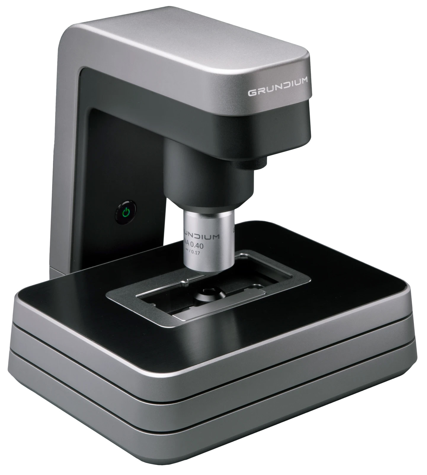
The Ocus®20 is a small, yet powerful microscope slide scanner for digital pathology, proven to be especially suitable for frozen section samples and rapid onsite evaluation (ROSE). It comes with a comparable magnification of 20x, meaning it is perfectly optimized for delivering sharp images fast.
SMALL FOOTPRINT
Most slide scanners are big pieces of equipment, requiring their own table or even room to operate, not always close to the lab. Because the Ocus®20 is so small, just 18 x 18 x 19 cm (ca. 8 x 8 x 8”), it can find a space in any corner of a desk in any lab. It weighs only 3,5 kg / 7,7 lbs, so it’s easy enough to just grab and take it with you, but also sturdy enough to be carried around without being damaged easily.
FAST AND EFFICIENT
For maximum throughput and speed multiple Ocus® scanners are run side by side, with one slide scanning while the next is being prepared. Read this testimonial about how Fimlab validated a fast digital frozen section process operated by laboratory technicians.
Scan time of a 15x15mm sample is circa 60 seconds. From Grundium’s browser-based user interface, the images can immediately be exported in JPG, SVS and TIFF formats and shared via email, uploaded to cloud services or exported to an external USB drive – or all at once. In addition, the device acts as a slide server: it has an internal storage of 500 gigabytes and a secure remote access can be set up. Grundium also offers an API to integrate the Ocus® scanners to your own workflow. Read more about the API here.
DIGITAL PATHOLOGY MADE SIMPLE
The browser-based user interface of the Ocus®20 is so simple and intuitive that anybody can be trained to use it in just 15 minutes. A lab tech can prepare and scan the slide onsite, and an expert can sign in and diagnose on their computer just as well right next to the scanner as in a different country. The long 5 um depth-of-field, continuous autofocus and Z-stacking of up to 5 layers all together make sure to reveal sharp details even in less than perfect samples. This makes the Ocus®20 excellent for tissue pathology.
The Ocus®20 is an incredibly practical tool for tissue pathology, second opinion and sharing scanned images online. Book a personal, non-binding and free of charge demo here.
FEATURES
Fully suited for remote operation, enables seamless telepathology
Fast scanning speed
High depth of field
Easy to use, anybody can use it with 15 minutes of training
Fits any workflow, supports all key file formats
Pairs with any device with internet connection (computer, laptop, tablet)
Robust design, very low maintenance
Small size, travels anywhere in its own carry-on case
Affordable, no recurring fees, no hidden costs
Fast scan speed, run several units parallel for very high throughput
Ocus®20 is CE marked according to EU 2017/746 regulation for in vitro diagnostic (IVD) medical devices. In the USA, Grundium Ocus®20 is for research use only (RUO) and not for use in diagnostic procedures.
APPLICATION AREAS
Pathology (frozen section, ROSE, etc.)
Clinical research
AI diagnosis
Marine sciences
Environmental research
Geology
Material sciences
TECHNICAL SPECIFICATIONS
Numerical aperture: 0.4
Resolution, microscope: 0.50 μm/pix
Resolution, overview: 10 μm/pix
Depth of field: 5 μm
Focusing: Fully electronic
Image sensor: 12 Mpix
Slide capacity: 1
Slide format: 75 x 25 mm
Scan speed, 15 x 15 mm: ~1 min
Connectivity: 1 GigE, 802.11 ac WLAN
Image formats: TIFF, SVS
Storage: 500GB SSD
W x D x H: 18x18x19 cm / 7x7x7,5 inches
Weight: 3,5 kg | 7,7 lbs
Illumination: Koehler LED